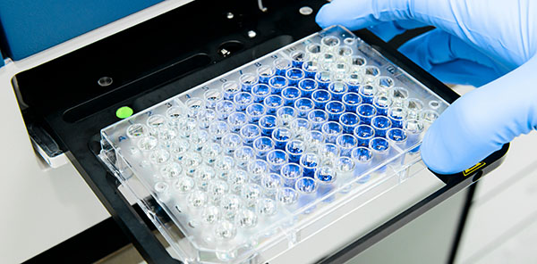
ELISA Troubleshooting
Several common issues can occur when running an ELISA. These are detailed in the following table, along with suggested solutions.
| No signal or weak signal | |
|---|---|
| Capture antigen or antibody may not have adhered to the microplate | |
| Check the binding capacity of the microplate according to the manufacturer’s description | |
| Try using a different coating buffer | |
| Increase the incubation time for plate coating | |
| Concentration of analyte-specific antibodies may be too low | Increase the concentration of the capture antibody and/or the analyte-specific detection antibody |
| Increase the incubation time for analyte-specific antibody binding | |
| Analyte-specific detection antibody and secondary antibody may be incompatible | Confirm that the host species of the analyte-specific detection antibody is compatible with the secondary antibody (for example, if the analyte-specific detection antibody was raised in rabbit, an anti-rabbit secondary antibody is required for detection) |
| Capture and detection antibodies may be competing for the same epitope (sandwich ELISA) | Confirm that the capture and detection antibodies recognize different epitopes |
| Switch to using a matched antibody pair if available | |
| Consider using a different ELISA format (e.g. try using an indirect ELISA that requires only one analyte-specific antibody rather than a sandwich ELISA) | |
| Fluorophore-conjugated antibodies may have been compromised by exposure to light | Ensure fluorophore-conjugated antibodies are stored correctly |
| Protect fluorophore-conjugated antibodies from light when adding them to, and incubating them in, the microplate wells | |
| If only the standard wells (and not the sample wells) are affected, the standard may have degraded | Try using a fresh vial of standard |
| Check that the standard has been prepared and stored correctly | |
| Azide (often added to antibody storage buffers as a preservative ) may be inhibiting HRP activity | Ensure sufficient washing to remove any residual traces of azide |
| Sample material may contain only low levels of the target analyte | Obtain more concentrated samples |
| Spike samples with a known amount of analyte to check the sample matrix is not a source of interference | |
| ELISA kits / kit components may have been stored incorrectly | Check the manufacturer’s instructions for storage |
| Plate may have been read at an incorrect wavelength | Check the reader settings are compatible with the chosen detection method |
| High Background | |
|---|---|
| Washing may be inadequate | Increase the number and/or duration of wash steps |
| Try adding detergent (e.g. 0.01-0.1% Tween-20) to wash buffers | |
| Blocking may be insufficient | Try using a more concentrated blocking solution |
| Increase the incubation time for blocking | |
| Switch to using a different blocking buffer | |
| Sample may be too concentrated | Try diluting the sample |
| Antibody concentration(s) may be too high | Decrease the concentration of the capture antibody, analyte-specific detection antibody, or secondary antibody |
| Decrease the antibody incubation time | |
| Colorimetric substrates may have been prepared too early | Always prepare substrates such as TMB immediately prior to use to avoid unwanted color development |
| Microplates may have sat around after the addition of stop solution (colorimetric detection) | Read colorimetric assays as soon as the stop solution has been added |
| Consumables such as pipette tips, reservoirs or buffers may have introduced contamination | Use fresh plasticware for each step |
| Prepare fresh buffers for each assay | |
| Incubation times may have been too long | Always follow the protocol and be consistent with reagent additions and timing |
| Poor reproducibility between plates | |
|---|---|
| Plates may have been coated unevenly | Ensure all solutions are thoroughly mixed before coating the plates |
| Seal plates after adding the coating solution to prevent evaporation; such effects can especially be noticeable in edge wells | |
| Check pipettes have been calibrated and are performing as expected | |
| Washing may be inadequate | Increase the number and/or duration of wash steps |
| Confirm wells are fully emptied between washes | |
| Wells may contain bubbles | Centrifuge microplates briefly prior to reading |
| Plate seals may be a source of cross-contamination | Always use fresh plate seals between incubations |
| Poor reproducibility between runs | |
|---|---|
| Reagents may have degraded | Prepare fresh reagents (including buffers) for each assay |
| Check that the standard has been prepared and stored correctly | |
| Samples may have been handled incorrectly | Always store and handle samples with care and avoid repeat freeze-thawing |
| Assay conditions may be inconsistent | Ensure all protocol steps are performed reproducibly |
| Always run ELISAs under stable environmental conditions | |
| Edge effects / plate drift | |
|---|---|
| The plate reader may be misaligned | Read the plate, then rotate it by 180o and read again; if the effect remains in the same position, the reader may need to be repaired by a qualified service engineer |
| Solutions may be cold | Ensure all solutions are at room temperature upon addition to the microplate unless otherwise stated in the protocol |
| Delays may have occurred during reagent addition | Prepare suitable quantities of reagents (including dead volumes) for the assay to avoid running out part-way across a plate |
| Volumes may be uneven across the microplate | Seal plates between reagent additions to prevent evaporation |
| Only use calibrated pipettes | |
| Plates may have cross-contaminated one another | Avoid stacking plates during incubations |
| Learn more: | Do more: |
|---|---|
| Colorimetric western blotting | Spectra Viewer |
| Chemiluminescence western blotting | Antibodies for signal enhancement |
| Fluorescent western blotting | |


