
Epidermal Blister Roof
Epidermal Blister Roof
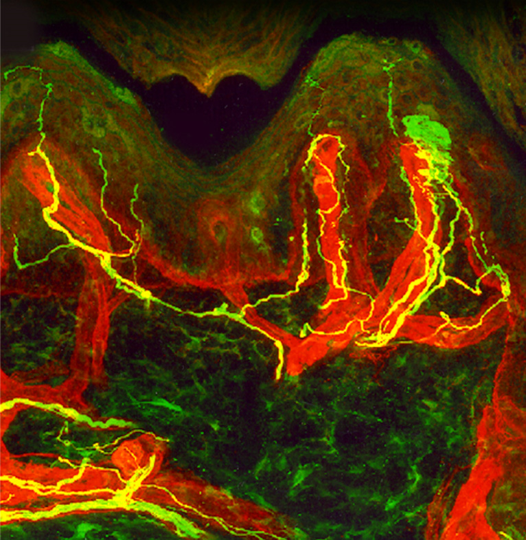
The epidermis was separated from dermis by creating a suction blister. The flattened blister roof was then processed without sectioning and stained for localization of nerve endings (Cy3, recolored green) and Langerhans cells (Cy2, recolored red) using Cy2- and Cy3- conjugated secondary antibodies from Jackson ImmunoResearch Laboratories, Inc. to provide a “bird’s eye view” of the distribution of nerve endings and dendritic cells within the epidermis. Some basal cells are weakly positive for the nerve marker.
| Product used: | Product code: |
|---|---|
| Cy3-AffiniPure Donkey Anti-Rabbit IgG (H+L) (min X Bov,Ck,Gt,GP,Sy Hms,Hrs,Hu,Ms,Rat,Shp Sr Prot) | 711-165-152 |
| Cy3-AffiniPure Donkey Anti-Mouse IgG (H+L) (min X Bov,Ck,Gt,GP,Sy Hms,Hrs,Hu,Rb,Rat,Shp Sr Prot) | 715-165-151 |
| Cy5-Streptavidin | 016-170-084 |
References:
Dr. William R. Kennedy and Dr. Gwen Wendelschafer-Crabb, Department of Neurology, University of Minnesota.
Technical Resources | About us | Contact us | Bulk Service
Licenses | Conditions of Use | Privacy Policy | Ordering Information

FM 545248