
Indirect immunofluorescence staining is a pivotal method for biomolecule labeling, yet its efficacy for super super-resolution microscopy can be hindered by large complexes that form between primary and secondary antibodies and fluorophores. This causes large linkage errors and artifacts due to the distance between the label and the target protein; therefore, solutions that increase the proximity between the fluorophore and the target molecule are crucial. We present a groundbreaking solution by combining abberior FLUX dyes with Jackson ImmunoResearch’s AffiniPure-VHH™, polyclonal VHH fragment secondary antibodies. Specifically optimized for single molecule localization techniques such as MINFLUX and STORM microscopy, abberior FLUX dyes ensure precise localization. Paired with JIR’s polyclonal VHH fragment antibodies (nanobodies) which bind to multiple sites of the primary antibody, they ensure remarkable signal amplification, resulting in captivating and informative images. This innovative combination heralds a new era in immunofluorescence labeling, offering superior performance, full flexibility, and enhanced visualization capabilities. Read our interview with abberior and Jackson ImmunoResearch on how these dyes and antibodies compliment each other…
Interviewees

Dave Fancy, Ph.D Jackson ImmunoResearch
Dave is the Chief Operating Officer for Jackson ImmunoResearch Laboratories. After gaining his Ph.D. from the University of Texas, Dave has specialized in the development of novel antibody conjugates of dyes, metal chelates, enzymes and DNA for use in the research. He has extensive experience in developing immunological products and services, with a focus on commercialization for the biotech research, diagnostic communities.

Evelyn Garlick, Ph.D., abberior Instruments
Evelyn comes from the UK and completed her Ph.D. at the Universities of Birmingham and Nottingham in 2022. Since then, she has been a part of the MINFLUX applications team at abberior Instruments, working to help users get the most out of their MINFLUX microscopes.

Florian Grimm, Ph.D., abberior dyes & labels
Florian, with a background in biochemistry and chemistry, was abberior’s first employee. With over a decade of experience with abberior dyes, labels, and microscopes, he now serves as a Business Developer. Florian is dedicated to finding innovative probes and maintaining key business relationships, driving abberior’s mission forward.
How does the format of AffiniPure VHH™ secondary antibodies help?
Fancy: The novel format of the VHH fragment is a 10th of the size of a conventional Ig antibody, this is good news if you want precise labeling where the fluorophore is close to your target protein because the primary/secondary complex is considerably reduced in size.

I’ve used monoclonal nanobodies, why should I use a polyclonal one?
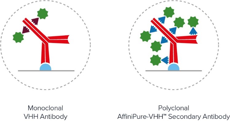
Fancy: The limitations of monoclonal antibodies are apparent when you need to achieve maximum brightness. The signal amplification achieved with polyclonal AffiniPure- VHH™ antibodies comes from the heterogenous population of antibodies, with different paratopes able to bind at many sites across the primary antibody ensuring it is maximally decorated with signal producing fluors. Figure x demonstrates the advantages of polyclonal VHH antibodies, binding across the whole of the primary antibody, rather than being limited to a specific site/region/sequence, enabling higher labeling efficiency and brighter signal than the monoclonal format.
I’ve got multiple targets; will these work together?
Fancy: JIR AffiniPure-VHH™ antibodies are cross-adsorbed against commonly used species to enhance specificity and thus reduce off-target labeling, they can be used in combination to generate exquisitely specific multiple labeling images.
What other benefits does the indirect method offer?
Fancy: Indirect labeling using conjugated AffiniPure-VHH™ Secondaries gives researchers access to a greater range of fluorophores, with AffiniPure VHH™ Secondaries available conjugated to fluors from ultraviolet to far-red researchers can pick the right characteristics for their experiments which is useful for those identifying multiple target proteins or seeking dyes suitable for SRM and SM techniques.
What is MINFLUX microscopy?
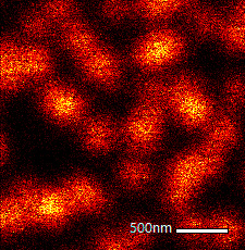
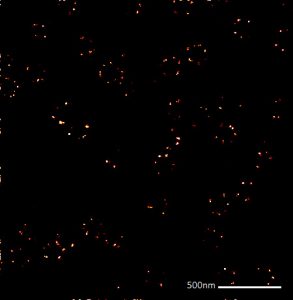
Garlick: MINFLUX (MINimal photon FLUXes) is a single molecule localization technique that finds fluorophores using an excitation minima rather than maxima. This difference is what makes MINFLUX the most precise and photon-efficient way of localizing fluorescent molecules, capable of achieving 1 – 3 nm localization precision in all three dimensions. The unparalleled precision afforded by MINFLUX nanoscopy has many applications, giving impressive insights into biological structures at the nanoscale.
What are FLUX dyes?
Grimm: FLUX dyes are organic fluorescent molecules that have been developed and tested for MINFLUX microscopy. The main feature of this particular class of dyes is that they start to blink through the use of a special buffer system. This blinking feature enables our dyes to oscillate between an ON and OFF state, a crucial aspect as it ensures that not all dyes are active simultaneously, allowing for precise localization of individual dye molecules over time.
But the versatility of abberior FLUX dyes doesn’t end there. They’re also compatible with other single-molecule localization microscopy techniques such as STORM, opening doors to many research possibilities.
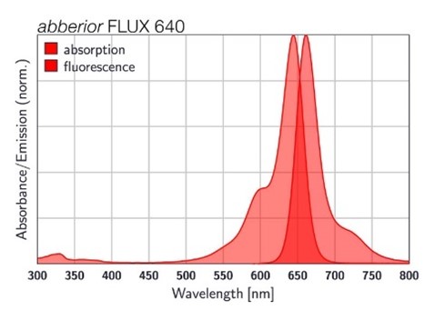
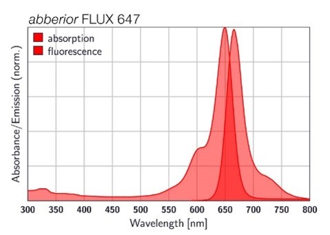
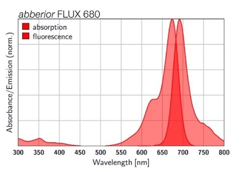
Figure 3: Absorption and fluorescence spectra of abberior FLUX dyes.
What is the advantage of FLUX dyes conjugated to AffiniPure-VHH™ antibodies?
Garlick: Dave explained that when using traditional primary and secondary antibodies, the distance from your epitope of interest to the fluorophore on your secondary can be ~ 20 nm. This is called linkage error. If you have a technique like MINFLUX that is capable of localizing a fluorophore with a precision of < 3 nm, reducing this linkage error is essential to better understand the arrangement of the epitope. AffiniPure-VHH™ Secondary antibodies can improve the linkage error, being much smaller than conventional Ig, they bring the fluorophore closer to the epitope, whilst keeping the flexibility of a traditional immunostaining approach.
How do i use abberior FLUX dyes conjugated to JIR AffiniPure-VHH Fragment antibodies?
Garlick: FLUX-conjugated VHH fragment secondary antibodies can easily be incorporated into existing staining routines in place of your conventional secondary antibody. A detailed protocol can be found here: https://abberior.rocks/expertise/protocols/nanobody-protocol/.
What is important when preparing a MINFLUX sample?
Garlick: Some special attention to your sample can help you achieve great quality MINFLUX results. In general, steps should be taken during labeling to limit background from both autofluorescence and unspecific labeling or binding. Make sure you incorporate a robust blocking and washing routine.
What dye to choose for single-color and which pair for two-color MINFLUX imaging?
Garlick: Our go-to dye recommendation for single-color imaging is FLUX 647. The best pair for two-color MINFLUX is FLUX 640 and FLUX 680 – these fluorophores are nicely spectrally separated in their emission and perform well in the same blinking buffer conditions, allowing simultaneous imaging of the two labels with the same excitation line.


| Learn more: | Do more: |
|---|---|
| Indirect and direct Western blotting | Exhibition Schedule |
| Chemiluminescence western blotting | Western blotting guide |
| An Introduction to Expansion Microscopy | ELISA guide |