According to Luminex® (a DiaSorin company), xMAP® is the world′s most used multiplexing technology, a claim that is backed by over 70,000 peer-reviewed publications. These include multiple references to serology assays developed using Jackson ImmunoResearch secondary antibodies, which serve as essential detection reagents for many diagnostic assays using this platform.
Principles of the Luminex® assay′
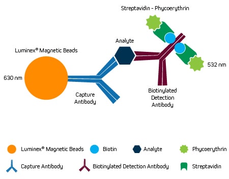
xMAP® Technology is based on the use of color-coded beads that are dyed with different ratios of two fluorophores (one red and one infrared) to provide a unique spectral signature. In a typical assay setup, each spectrally distinct bead set is coated with a different analyte-specific antibody and is initially blocked prior to incubation with the sample. After washing to remove any unbound material, biotinylated analyte-specific antibodies are added to form a sandwich complex before a streptavidin-phycoerythrin (PE) conjugate completes the assemblage. The beads are then read on a flow cytometry-based Luminex® instrument, where a red (630 nm) laser is used to identify each bead set and a green laser (532 nm) determines the magnitude of the PE-derived signal, which is directly proportional to the amount of bound analyte. This setup is shown in Figure 1.

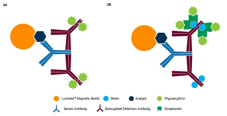
An alternative approach involves coating each bead set with a protein analyte to capture antibodies from sample types including serum, saliva, and cerebrospinal fluid. In this setup, detection is usually accomplished using a PE-conjugated anti-species antibody (Figure 2a) or a biotinylated anti-species antibody in combination with a streptavidin-PE conjugate. Secondary antibodies from Jackson ImmunoResearch are widely used for these types of assays.

Advantages of xMAP® Technology
Like other multiplexed immunoassay formats, xMAP® Technology offers savings in terms of sample material, reagents, and time, as well as eliminates confounding variables like different buffer systems, ambient conditions, and operators for a more accurate comparison of different analytes within the same assay. However, a distinguishing feature of xMAP® Technology is that it can simultaneously detect up to 500 analytes per sample, which is more than most other multiplexed immunoassays. Additionally, it is now possible with the xMAP INTELLIFLEX® to measure two parameters per analyte. “The xMAP INTELLIFLEX® enhances the capabilities of the original xMAP technology by incorporating a second (405 nm) detection channel,” explains Dominic Andrada, Scientific Applications Senior Marketing Manager at Luminex. “This enables researchers to effectively double the number of readouts obtained per sample. For example, using the xMAP INTELLIFLEX®, one can detect a protein of interest and a specific post-translational modification on the same bead.” Another important advantage of xMAP® Technology is that the small size and low density of the beads equates to shorter assay times than ELISAs and microarrays, which are limited by solid-phase kinetics1.
Applications of xMAP® Technology
Because xMAP® Technology is highly flexible, it has utility for an almost infinite number of research applications. These include its use to study cancer (e.g., through measuring immune checkpoint proteins, circulating tumor markers, or markers of metastasis), inflammation (e.g., by quantifying cytokines, chemokines, or autoantibodies), and cell signaling pathways (e.g., via monitoring of Akt/mTOR, NFκB, or STAT signaling molecules). xMAP® Technology is also used for investigating the underlying mechanisms of metabolism, aging, and virology, as well as for studying neurodegeneration, metabolic disorders, and cardiovascular disease.
High-quality reagents from Jackson ImmunoResearch for xMAP® assays
Jackson ImmunoResearch specializes in producing secondary antibodies for life science applications and our products have been extensively cited for use with xMAP® Technology.
Examples include
- 2021 Scientific Reports publication in which our biotinylated donkey anti-human IgG Fc-specific antibody was used for detecting IgG antibodies to three noroviruses and Cryptosporidium in the saliva of individuals who reported swallowing water from Lake Michigan.
- 2023 Immunology article citing our PE-labeled goat anti-human IgG antibody for detecting anti-SARS-CoV-2 antibodies in serum2,3.
- R-Phycoerythrin AffiniPure F(ab′)₂ Fragment Goat Anti-Mouse IgM, µ chain specific and R-Phycoerythrin AffiniPure F(ab′)₂ Fragment Goat Anti-Mouse IgG, Fcγ fragment specific antibodies featured in an SLAS publication reporting the development of an xMAP® assay for detecting anti-glycan antibodies in plasma, ascites, and cerebrospinal fluid4.
Changes in glycosylation have been linked to congenital disorders, autoimmune diseases, and cancer, and it is thought that generating anti-glycan antibody profiles from different human body fluids may help in predicting disease outcomes or patient responses to treatment.
References:
- Khalifian, S., Raimondi, G., & Brandacher, G. (2015). The use of luminex assays to measure cytokines. The Journal of investigative dermatology, 135(4), 1–5. https://doi.org/10.1038/jid.2015.36/
- Egorov, A. I., Converse, R., Griffin, S. M., Bonasso, R., Wickersham, L., Klein, E., Kobylanski, J., Ritter, R., Styles, J. N., Ward, H., Sams, E., Hudgens, E., Dufour, A., & Wade, T. J. (2021). Recreational water exposure and waterborne infections in a prospective salivary antibody study at a Lake Michigan beach. Scientific reports, 11(1), 20540. https://doi.org/10.1038/s41598-021-00059-2/
- Lundin, S. B., Kann, H., Fulurija, A., Andersson, B., Nakka, S. S., Andersson, L. M., Gisslén, M., & Harandi, A. M. (2023). A novel precision-serology assay for SARS-CoV-2 infection based on linear B-cell epitopes of Spike protein. Frontiers in immunology, 14, 1166924. https://doi.org/10.3389/fimmu.2023.1166924/
- Antonia Katharina Hefermehl, Sanne Maria Mathias Hensen, Carina Versantvoort, Andrée Rothermel, Uğur Şahin, Automated glycan-bead coupling for high throughput, highly reproducible anti-glycan antibody analysis, SLAS Technology,2023,,ISSN 2472-6303, https://doi.org/10.1016/j.slast.2023.08.003.5
- Luminex® and xMAP INTELLIFLEX® are registered trademarks of Luminex Corporation.
| Learn more: | Do more: |
|---|---|
| Colorimetric western blotting | Spectra Viewer |
| Chemiluminescence western blotting | Antibodies for signal enhancement |
| Fluorescent western blotting | |


