.
Multiplexed immunoassays offer many advantages. These include more data points per sample, higher throughput, and (in many cases) reduced cost. Multiplexing also eliminates confounding variables such as assay temperature, buffer system, and operator by measuring all of the analytes under identical experimental conditions at the same time. A main challenge of developing multiplexed immunoassays is that the concentration and expression pattern of different analytes can vary significantly. This can be addressed through careful experimental design.

1. Understand your analytes
For a multiplexed immunoassay to generate reliable data, it is critical that it be configured to ensure accurate detection of each of the analytes in question. Analytes can differ in abundance as well as in where they are expressed; while some analytes are intracellular, others are expressed at the cell surface or as secreted biomolecules. In certain cases, it may be necessary to stimulate analyte expression (e.g., by incubating with a growth factor) or to identify a specific protein isoform. Open-access resources such as UniProt and PAXdb offer information about protein abundance, known isomers, and post-translational modifications (PTMs), while publications and antibody datasheets are other useful resources.
2. Consider the level of multiplexing required
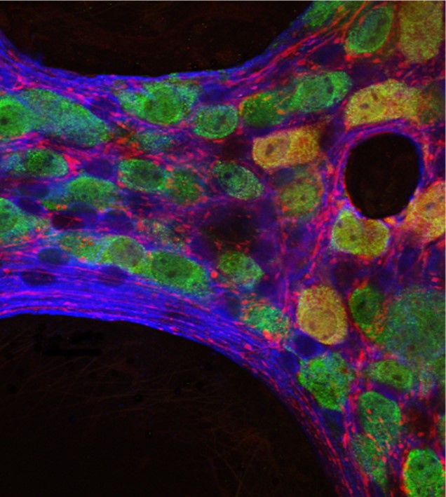
Multiplexed immunoassays range in complexity, spanning Western blot detection of a target protein and a loading control on the same membrane, through immunohistochemical (IHC) analysis of 3 or 4 distinct proteins in a tissue section, to flow cytometry-based detection of as many as 20 different analytes expressed by single cells. Using bead-based assays or antibody arrays, it is possible to monitor many hundreds of biomolecules simultaneously. As a general rule, the less that is known about a particular analyte, the higher the level of multiplexing that is required; although it can be tempting to detect as many targets as possible, the number of analytes should be balanced against the cost per test and the amount of work involved in assay development and validation.
3. Identify suitable analyte-specific antibodies
Analyte-specific antibodies are critical components of a multiplexed immunoassay and should be selected with care. First, they should be validated for the intended application, with the antibody manufacturer’s datasheet providing supporting evidence for antibody performance. Equally important is the need to titrate antibodies to identify an acceptable signal window; this includes both analyte-specific antibodies and any secondary antibodies that will be used for their detection. Monoclonal antibodies offer the advantages of consistent batch-to-batch performance and guaranteed long-term supply. Yet, because polyclonals can provide signal amplification by recognizing several epitopes, they can be a preferred choice for detecting rare analytes. It is common practice for both monoclonal and polyclonal antibodies to be combined in the same experiment for multiplexed detection.
4. Select compatible secondary antibodies
Unless analyte-specific antibodies are available conjugated to enzymes or fluorophores, secondary antibodies are essential reagents for enabling target detection. Importantly, secondary antibodies can provide signal amplification, meaning they are often incorporated into multiplexed immunoassay workflows to ensure low abundance targets are detected. When selecting secondary antibody reagents, it is vital to confirm they are compatible with analyte-specific antibodies – for example, a goat anti-rabbit secondary would be used to detect an analyte-specific antibody raised in a rabbit host. Subclass-specific secondary antibodies offer the flexibility to use several antibodies from the same host species in parallel, provided their specificity has been confirmed by the manufacturer, whereas cross-adsorbed secondary antibodies (Min X) minimize species cross-reactivity that may be a source of unwanted background signal. There are also strategies for labeling two primary antibodies from the same host species when other primary antibody options are not available here.
5. Determine whether additional signal amplification is necessary
Although polyclonal analyte-specific antibodies and secondary antibody reagents both provide signal amplification, it can sometimes be necessary to boost the assay signal further still. A popular approach is to use labeled streptavidin-biotin (LSAB) signal amplification, whereby the analyte-specific antibody is bound by biotinylated secondary antibodies before the addition of labeled streptavidin for detection. In this situation, signal amplification is achieved through 1) multiple biotins being attached to each secondary antibody and 2) the tetrameric nature of streptavidin, which has four biotin-binding sites per molecule increases avidity.
6. Use online tools to simplify panel design
In the majority of cases, multiplexed immunoassays rely on fluorescent readouts (an exception is Western blotting, where chemiluminescent detection may be used to visualize two bands with distinct molecular weights simultaneously) and it is advised that bright fluorophores be paired with rare targets, and vice versa. As the number of target analytes increases, panel design becomes more challenging, especially for flow cytometry where as many as 20 different biomolecules may be detected in the same experiment. Online tools such as Spectra Viewer simplify panel design by providing visualization of fluorophore excitation and emission spectra, enabling rapid assessment of fluorophore compatibility.
7. Remember to confirm batch-to-batch consistency
Multiplexed immunoassays involve using a broad selection of antibodies, each of which will comprise different batches over time. It is recommended that researchers test antibody performance every time a new batch is purchased – including both analyte-specific antibodies and secondary antibody reagents – to ensure reproducible results. For studies involving large sample numbers, or those that will run over an extended period of time, it can be sensible to purchase (or reserve) bulk quantities of antibody reagents directly from the manufacturer.
8. Always include appropriate controls
Controls are essential to any immunoassay, where they are used to monitor assay performance and validate results. Positive controls produce a maximal assay signal, while negative controls provide an indication of non-specific binding; both should be included in every immunoassay, irrespective of the number of different targets being detected. Other types of controls include secondary antibody-only controls that are designed to evaluate secondary antibody binding in the absence of analyte-specific antibodies; compensation controls that are used to assess spectral overlap in flow cytometry by staining samples with a single fluorophore; and isotype controls (comprising an antibody of the same isotype and concentration as the analyte-specific antibody but which does not recognize the target) that are used in both IHC and flow cytometry to help identify sources of background staining.
9. Think about incorporating automation
Automation complements the benefits of multiplexing by increasing sample throughput and improving experimental consistency. It also reduces the risk of cross-contamination, helps prevent repetitive strain injuries to body, and frees up researchers’ time to be spent on other tasks. Automation options include multichannel dispensers that can dispense 96 aliquots simultaneously, IHC staining systems, and fully automated immunoassay platforms; these are all worth considering as strategies for streamlining multiplexed workflows.
10. Never hesitate in asking for help
Setting up a multiplexed immunoassay can be daunting, but Jackson ImmunoResearch – a specialized producer of secondary antibodies for life science applications – is here to help. We offer a broad range of secondary antibodies conjugated to fluorescent proteins (including R-PE, APC, and PerCP) and fluorescent dyes (including Alexa Fluor®, Brilliant Violet, and Rhodamine Red™), as well as subclass-specific secondary antibodies, cross-adsorbed secondary antibodies, biotinylated secondary antibodies, and labeled streptavidin. To streamline panel design, researchers can access our Spectra Viewer, while our online technical support options include a comprehensive secondary antibody resource featuring protocols and technical tips, and a broad range of guides for product selection and use.
For more information on experimental design and choosing reagents for immunofluorescence multiple labeling see here.
| Learn more: | Do more: |
|---|---|
| Colorimetric western blotting | Spectra Viewer |
| Chemiluminescence western blotting | Antibodies for signal enhancement |
| Fluorescent western blotting | |


