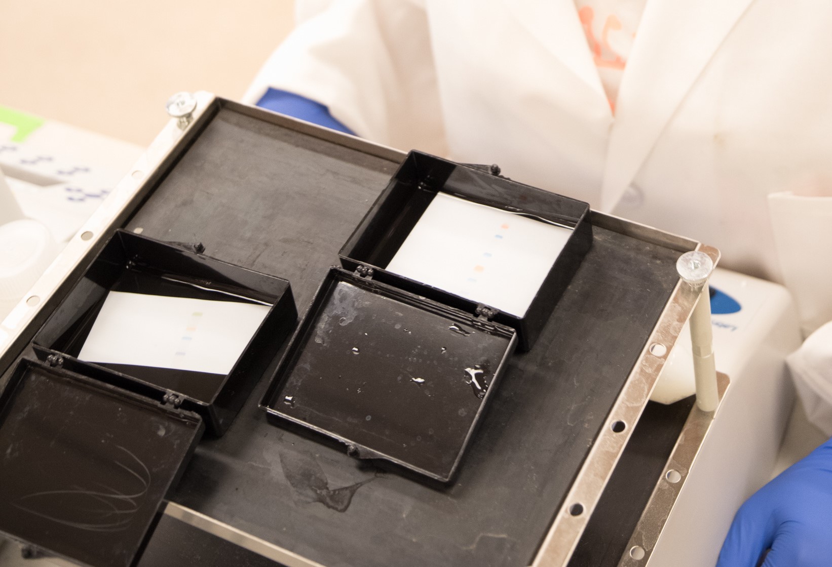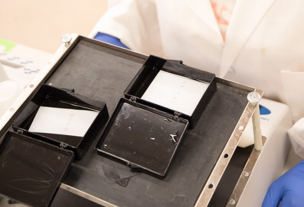
Blocking is essential to prevent non-specific interactions between the transferred proteins, the membrane and the subsequent reagents used in an experiment. Non-specific binding can lead to background signal, which can then interfere with result interpretation. In Part 4 of our Western blotting guide we detail the correct way to block and prevent unwanted non-specific interactions.

To prevent any non-specific interactions between the transferred proteins, the membrane and the subsequent reagents used in an experiment, the membranes are blocked before they are probed with antibodies. Non-specific antibody-membrane binding can cause background signal, which can then potentially interfere with result interpretation (such as the misidentification of the target protein(s)).
Ideally, an optimal blocking buffer “should improve the sensitivity of the WB by reducing background and improving the signal-to-noise ratio (measured as the signal obtained in a sample containing the target analyte vs. obtained with a sample without the target analyte).”4
Blocking is achieved by combining a protein-rich solution with other reagents, such as non-ionic detergents, to saturate any sites on the membrane that could interact with antibodies used to detect and visualize the protein of interest and optimize the signal-to-noise ratio. The blotted membrane is immersed in a freshly prepared blocking solution for a minimum of one hour at room temperature (although extended incubation periods may be required to optimize blocking efficiency) and placed on a rocker or shaking platform.
Choosing an Appropriate Blocking Reagent
The choice of the blocking reagent is indicated by the species the secondary antibody used in the detection step of the western blot.
Protein Blocking Reagent
The protein component of the blocking solution should be compatible with both the antigen that is being detected and the species of the secondary antibody.
Bovine serum albumin (BSA) and non-fat-dried milk are commonly used in blocking solutions as they are both inexpensive and easily available. However, they are not always appropriate and can be a cause of background.
BSA and non-fat-dried milk should be avoided when using antibodies directed against antibodies derived from bovine or related species such as alpaca, goat, horse, or sheep. Low levels of bovine immunoglobulin G (IgG) are found in many BSA formulations.
Normal serum from the host species of the labeled secondary antibody is recommended as the best blocking agent to avoid any unwanted interactions. A fresh working solution of serum should be prepared before use and is typically diluted to 5% w/v in the chosen buffer before the membrane is immersed.
Buffer Solutions
Buffer solutions with pH ranges higher than the isoelectric point (pI) of most antibodies (pH range 7.5-8.5), such as Tris-buffered saline (TBS) or phosphate-buffered saline (PBS), are often used. PBS is not recommended if phosphorylated proteins are the focus of the blot.
This is because the primary antibody can bind to phosphate in the buffer as well as the target protein. Buffer pH should also be considered for its effect on the activity of reporter enzymes used in the detection step (e.g., alkaline phosphatase).
Non-Ionic Detergents
Non-ionic detergents are often included in buffers used in Western Blotting. They prevent aggregation, which in itself can cause background, but also binding to any sites which might cause non-specific interactions.
Tween-20™ (typically 0.1%) is commonly used as a buffer additive.
Incubation Duration and Washing
The shortest possible duration of blocking required to achieve an optimal signal-to-noise ratio should be determined. Although overnight blocking at 4°C may be attractive, overextending blocking incubations can lead to aggregation and bacterial growth, which may cause background or interfere with visualization.
The blocking process may require optimization by varying the components and their concentration, time, and temperature to obtain the lowest background and highest signal-to-noise ratio.
Notably, washing the membrane after blocking with a buffer without its protein component is important because it may improve access of the primary antibody to the antigenic epitope.
|
Blocking Reagent |
Concentration |
Contraindication |
|---|---|---|
|
Non-fat (skimmed) milk powder |
3-5% w/v |
Do not use dried milk to block when using anti-phospho antibodies as the casein in milk (a phospho protein) will interact. Do not use BSA or dried milk with secondary antibodies raised against alpaca, goat, sheep, horse, or cow. Do not use milk when using Biotin/avidin/streptavidin in later steps as milk contains biotin, so would interfere with biotin-avidin/streptavidin detection systems. |
|
BSA |
2-10% w/v |
Do not use BSA if it was used during the immunization process to generate antibodies. Do not use BSA or dried milk with secondary antibodies raised against alpaca, goat, sheep, horse, or cow. |
|
Normal serum from the species of the labeled antibody |
5% w/v |
Note: If normal serum from the species in which the PRIMARY antibody was made is used by mistake, significant background will result! |
|
PBS |
|
Do not use phosphate buffer if using Alkaline phosphatase as a conjugate. |
Caution: proprietary blocking solutionsSeveral preformulated blocking solutions are available, which are marketed as suitable for all experimental conditions. This may not always be the case, although they may be adequate for one set of conditions. It is important to consider the species of subsequent reagents may preclude the use of generic blocking solutions, and using the normal serum of the species of the labeled antibody may be more reliable. |
Caution: Blocking with BSA or milkBovine serum albumin (BSA) and dry milk can contain bovine IgG. Anti-Alpaca, Anti-Bovine, Anti-Goat, Anti-Horse and Anti-Sheep antibodies will react with bovine IgG. Use of BSA or dry milk for blocking or diluting may significantly increase background and/or reduce antibody titer and should be avoided for antibodies with these specificities; use the normal serum (5% v/v) of the host species of the labeled secondary antibody. If BSA is being considered as a blocking agent, use those that are IgG-free to reduce background potential. |
|
Problem |
Cause |
Solution |
|---|---|---|
|
Background (non-specific signal obscuring bands of interest) |
Improper blocking.
Use appropriate blocking reagent to block the membrane before incubating with the primary antibody.
Avoid using milk or BSA if using primary antibodies derived from goat, horse, or sheep. Bovine IgG may interact with the antibody due to homologous epitopes of closely related species. |
Extend the duration of blocking incubation.
Normal serum (5% v/v) from the host species of the labeled antibody, or BSA (5% w/v) (IgG-free and protease-free).
Normal serum (5% v/v) from the host species of the labeled antibody. |
|
High Background/low-signal-to-noise ratio. |
Low blocking concentration |
Use a negative control to check that the primary antibody detects the target specifically. |
|
Poor signal |
Blocking reagent masking epitope from the primary antibody. Non-specific primary antibody
|
Decrease blocking agent concentration, reduce incubation length, choose a different blocking reagent, or add a wash step after blocking. Use a positive control to check the specificity of the primary antibody. (see primary antibody section). |
Found Part 4 of our Western blotting guide useful? View part 5 here
References
- Blancher C. et al. (2001). SDS-PAGE and Western Blotting Techniques. Methods in Molecular Medicine. doi: 10.1385/1-59259- 136-1:145.
- Jackson ImmunoResearch Laboratories Inc. (2017). Western Blotting Troubleshooting Guide! https://www.jacksonimmuno.com/secondary-antibody-resource/technical-tips/western-blot-trouble-shooting/
- GE Healthcare. (2011). Western Blotting Principles and Methods. https://www.sigmaaldrich.com/content/dam/sigma-aldrich/docs/Sigma-Aldrich/General_Information/1/ge-western-blotting.pdf
- Bass J.J. et al. (2017). An Overview of Technical Considerations for Western Blotting Applications to Physiological Research. Scandinavian Journal of Medicine and Science in Sports. doi: 10.1111/sms.12702
- Lewis M. (2018). A Guide to Selecting Control and Blocking Reagents. Jackson ImmunoResearch Laboratories Inc. https://www.jacksonimmuno.com/secondary-antibody-resource/technical-tips/controls-diluents-blocking/
- Kelly M.F. et al. (2016). Antibody Validation. Mater Methods. doi.org/10.13070/mm.en.6.1540
- Lewis M. (2018). Cross-Adsorbed Secondary Antibodies and Cross-Reactivity. Jackson ImmunoResearch Laboratories Inc.https://www.jacksonimmuno.com/secondary-antibody-resource/technical-tips/cross-adsorbed-and-cross-reactivity/
- https://www.birmingham.ac.uk/Documents/college-mds/facilities/crf/scientists/western-blotting.pdf
| Learn more: | Do more: |
|---|---|
| Colorimetric western blotting | Spectra Viewer |
| Chemiluminescence western blotting | Antibodies for signal enhancement |
| Fluorescent western blotting | |


