
Imaging using immunostaining is a commonly used technique that can elucidate many details about how a process functions in situ. It is applied in many fields, including cancer and neuroscience research, to create a better understanding of biological processes. Although immunostaining is a robust and straightforward proposition, as the complexity of the experiment increases to dissect more complicated questions, experimental design requires reagents that enable specific detection. The following article introduces how Jackson ImmunoResearch cross-adsorbed antibodies and NEW Anti-GFP antibodies, can benefit your research.
Cross-adsorbed antibodies for exquisite differentiation
Secondary antibodies raised against one species may recognize epitopes on other species’ immunoglobulins through structural similarities, known as homology. This cross-reactivity can be problematic and cause background tissue staining or signal by recognizing a non-target primary antibody. When experiments combine material from multiple species, such as presented by sample species or multiple primary antibodies presenting different hosts, secondary antibodies with minimal cross-reactivity (min X) to serum proteins from non-target species are necessary to avert background from cross-reactivity.
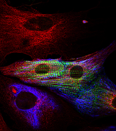
Determining Cardiomyocytes from heart primary cultures: 3 cell types are identified in this image. Red only cells show fibroblasts (positive for Rat Anti-tubulin); Smooth muscle cells are positive for both tubulin (red) and desmin (blue). Cardiomyocytes are identified in this image by their exhibition of green stripes, positive for MyBP-C.
To find out more about Dr Ehler’s research read here.
Dr Elisabeth Ehler, King’s College London
Cross-absorbed antibodies from Jackson ImmunoResearch are designated as “(min X … Sr Prot)” in their product description. These antibodies exhibit minimal cross-reactivity with the species detailed in the parenthesis, whether presented by the experimental antibodies or by endogenous proteins in the sample. For example, AffiniPure® Donkey Anti-Mouse IgG (H+L) (min X Bov, Ck, Gt, GP, Sy Hms, Hrs, Hu, Rb, Rat, Shp Sr Prot) antibody has been cross-adsorbed against bovine, chicken, goat, guinea pig, Syrian hamster, horse, human, rabbit, rat and sheep IgG and serum proteins and is suitable for experiments where those species are used. You can learn more about multiple labeling and cross-adsorbed antibodies by following the links below.
Anti-GFP Antibodies for the detection of GFP proteins
Jackson ImmunoResearch AffiniPure™ Rabbit Anti-GFP is an affinity-purified polyclonal antibody raised in rabbit and is specific for Aequorea victoria Green Fluorescent Protein (avGFP) and its derivatives, including EGFP, ECFP, and EYFP. These antibodies are versatile reagents and may be used in a wide range of applications, including ELISA, IHC, ICC, IP, Flow cytometry, and WB.
Imaging with Anti-GFP
Conjugates for immunofluorescence
This Anti-GFP antibody is available conjugated to a range of reporter molecules, including Alexa Fluor® dyes to enable indirect detection for bright target signal and spectacular signal amplification.
They enable circumvention of autofluorescence or equipment limitations, such as when a laser line or channel is occupied by another target, making them useful when detecting multiple targets in the same experiment.